
A comprehensive shoulder examination evaluates the structure and function of the shoulder joint, muscles, and tendons. It identifies common pathologies like rotator cuff injuries and instability, guiding diagnosis and treatment planning effectively.

Essential Components of Shoulder Examination
A thorough shoulder exam includes patient history, visual inspection, palpation, range of motion assessment, strength testing, and special tests to diagnose injuries or conditions effectively.
2.1 Patient History and Questionnaire
A patient history and questionnaire are critical for identifying symptoms, onset, and aggravating factors. They provide insights into pain characteristics, past injuries, and lifestyle, guiding the physical examination. Standardized questions help assess functional limitations and prior treatments, ensuring a comprehensive understanding of the condition. This step also identifies red flags for serious conditions, such as sudden trauma or neurological symptoms, ensuring timely referrals. A detailed history enables clinicians to tailor the exam and diagnostic process effectively, improving accuracy in identifying shoulder pathologies. It is the foundation of a systematic shoulder evaluation.
2.2 Inspection
Inspection begins with a visual assessment of the shoulder, noting posture, muscle atrophy, swelling, or deformities. The clinician observes for asymmetry, scapular positioning, and signs of inflammation. The patient is examined from multiple angles to identify any visible abnormalities. This step helps detect conditions like shoulder dislocation or impingement; The presence of scars or redness may indicate prior trauma or surgery. Inspection provides a baseline for further examination and helps prioritize subsequent tests, ensuring a systematic approach to identifying shoulder pathologies. It is a non-invasive yet crucial initial step in evaluating shoulder health and function.
2.3 Palpation
Palpation involves manually examining the shoulder to assess tenderness, swelling, or abnormalities. The clinician gently presses on key areas, including the clavicle, acromion, rotator cuff muscles, and joint capsule. This step helps identify localized pain, muscle spasms, or structural irregularities. Palpation is crucial for detecting issues like rotator cuff tears or bursitis. The examiner uses fingertips to apply controlled pressure, ensuring a thorough yet gentle assessment. This technique complements inspection by providing tactile feedback, aiding in the diagnosis of underlying shoulder pathologies and guiding further clinical evaluation.
2.4 Range of Motion Assessment
Range of motion assessment evaluates the shoulder’s mobility in flexion, extension, abduction, internal rotation, and external rotation. Active movements are performed by the patient, while passive movements are guided by the examiner. This step identifies limitations or pain during specific motions, which may indicate injuries or conditions like impingement or frozen shoulder. Documentation of reduced or painful mobility aids in diagnosing pathologies and developing targeted rehabilitation plans. Accurate measurement using a goniometer ensures objective data collection, crucial for monitoring progress and treatment effectiveness.
2.5 Strength Testing
Strength testing evaluates the shoulder muscles’ power and endurance, focusing on key muscle groups like the supraspinatus, deltoids, and rotator cuff. Manual resistance or dynamometers measure strength, identifying weaknesses or imbalances. This assessment helps diagnose conditions such as rotator cuff tears or muscle strains. By comparing bilateral strength, examiners can detect unilateral deficits, guiding rehabilitation strategies. Objective measurements aid in monitoring progress and ensuring effective treatment plans.
2.6 Special Tests
Special tests in shoulder examination are designed to assess specific pathologies. The Neer test and Hawkin’s test identify impingement syndrome, while the apprehension test evaluates anterior instability. The drop arm test detects supraspinatus tears, and the sulcus test checks for inferior instability. These maneuvers provoke symptoms or demonstrate anatomical disruptions, aiding in accurate diagnosis. They are essential for differentiating between various shoulder conditions and guiding further investigation or treatment plans.
Clinical Relevance of a Shoulder Examination
A thorough shoulder examination is crucial for diagnosing and managing shoulder-related conditions; It enables early detection of pathologies, such as rotator cuff injuries or instability, which are common causes of pain and dysfunction. Accurate assessment informs treatment plans, reducing the risk of chronic issues or unnecessary surgical interventions. By identifying specific limitations and impairments, clinicians can tailor rehabilitation strategies, improving patient outcomes. Regular monitoring of shoulder function also aids in tracking recovery progress, ensuring effective management of both acute and chronic conditions.

Common Shoulder Pathologies
Common shoulder pathologies include rotator cuff injuries, shoulder instability, and impingement syndrome; These conditions often cause pain, limited mobility, and functional impairment, requiring precise diagnostic and therapeutic approaches.
4.1 Rotator Cuff Injuries
Rotator cuff injuries are among the most common shoulder pathologies, often caused by overuse, trauma, or degeneration. Symptoms include pain, weakness, and limited arm mobility. Partial or full-thickness tears in the tendons or muscles of the rotator cuff can significantly impair function. Early diagnosis through physical examination and imaging is crucial for effective treatment. Conservative management includes physical therapy, anti-inflammatory medications, and activity modification. Severe cases may require surgical intervention to repair the damaged tissues. Proper rehabilitation is essential to restore strength and mobility, preventing further complications and improving quality of life.
4.2 Shoulder Instability
Shoulder instability refers to the excessive movement or displacement of the shoulder joint, often due to trauma, repetitive strain, or congenital factors. It can be traumatic or atraumatic, with symptoms including pain, apprehension, and limited mobility. Diagnosis involves physical examination maneuvers and imaging studies like X-rays or MRIs. Conservative treatment includes physical therapy to strengthen stabilizing muscles, while severe cases may require surgical intervention to restore joint stability. Early identification and appropriate management are crucial to prevent recurrent dislocations and maintain optimal shoulder function.
4.3 Impingement Syndrome
Impingement syndrome occurs when the rotator cuff tendons or bursa become compressed under the acromion, leading to pain and limited shoulder movement. Common causes include repetitive overhead activities, bone spurs, or poor posture. Symptoms often worsen with abduction or internal rotation. Diagnosis involves clinical tests like the Hawkins-Kennedy test and imaging such as X-rays or MRIs. Conservative management includes physical therapy to strengthen the rotator cuff and improve posture, as well as anti-inflammatory medications. Severe cases may require arthroscopic surgery to relieve compression and restore normal shoulder function.

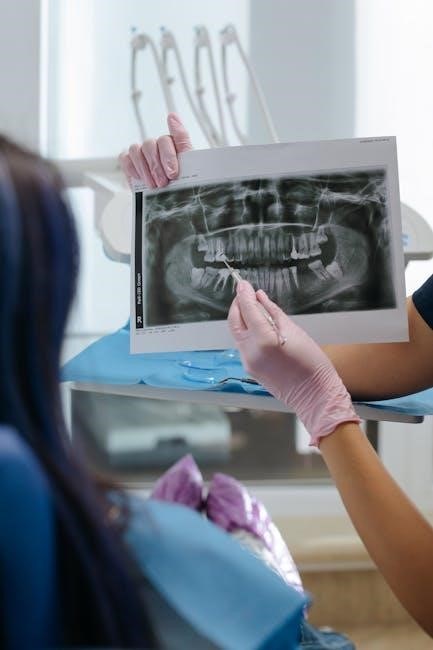
Diagnostic Imaging in Shoulder Examination
Diagnostic imaging plays a crucial role in evaluating shoulder injuries and pathologies. Common imaging modalities include X-rays, MRI, CT scans, and ultrasound. X-rays are often the first-line imaging method to assess bone fractures or degenerative changes. MRI provides detailed images of soft tissues like the rotator cuff, tendons, and labrum, making it ideal for diagnosing injuries or inflammation. CT scans are useful for complex fractures or surgical planning. Ultrasound offers real-time visualization of dynamic shoulder movements and is beneficial for guiding injections or assessing tendons and ligaments. These imaging tools help confirm clinical findings and guide appropriate treatment strategies.

Rehabilitation and Management Strategies
Effective rehabilitation focuses on restoring shoulder function, strength, and mobility. Physical therapy is cornerstone, incorporating exercises for range of motion, strengthening, and proprioception. Pain management may include NSAIDs or corticosteroid injections. Activity modification is essential to avoid aggravating movements. A structured program often progresses from passive to active exercises, emphasizing proper technique. Patient education on posture, ergonomics, and injury prevention is vital. Rehabilitation aims to enhance quality of life and return patients to normal activities, ensuring long-term shoulder health and functionality.
Documentation and Reporting
Accurate documentation is critical for effective communication and continuity of care. Detailed records should include patient history, examination findings, diagnoses, and treatment plans. Standardized templates can ensure consistency. Progress notes should track improvements or setbacks, guiding further interventions. Clear documentation also serves as a legal record of interactions and decisions. Reports should be concise, yet comprehensive, facilitating collaboration among healthcare providers. Proper documentation ensures transparency, accountability, and optimal patient outcomes, making it a cornerstone of professional practice in shoulder examination and management.

Physical Examination Techniques
Physical examination techniques involve systematic inspection, palpation, and assessment of range of motion to evaluate shoulder structure and function, aiding in the identification of potential pathologies.
8.1 Hands-On Methods
Hands-on methods are crucial in shoulder examinations, involving palpation, range of motion assessment, and strength testing. Palpation helps identify tenderness in the rotator cuff and acromioclavicular joint. Passive and active range of motion tests evaluate flexibility and mobility. Strength testing, such as the supraspinatus test, assesses muscle function. Special tests like the Hawkins-Kennedy test for impingement and the Apprehension test for instability provide diagnostic clarity. These techniques allow clinicians to accurately identify structural and functional abnormalities, guiding further diagnostic or therapeutic interventions effectively.
8.2 Observation Techniques
Observation is the first step in shoulder examination, providing valuable insights into potential pathologies. Clinicians assess posture, muscle symmetry, and visible deformities. Scapular positioning, atrophy, or swelling may indicate underlying issues. Comparative analysis of both shoulders helps identify asymmetries. Observing active movements, such as abduction and rotation, reveals limitations or compensatory patterns. This visual assessment guides further hands-on evaluation and helps prioritize diagnostic tests, ensuring a comprehensive understanding of the shoulder’s functional and structural status.

Red Flags and Referral Criteria
Red flags in shoulder examination include sudden weakness, numbness, or severe pain, which may indicate neurological or vascular issues. Systemic symptoms like fever or unexplained weight loss suggest underlying conditions requiring further investigation. A history of trauma, especially in older adults, warrants imaging to rule out fractures. Persistent pain or limited mobility despite conservative treatment may necessitate specialist referral. Early identification of these flags ensures timely intervention, preventing complications and improving outcomes for patients with shoulder-related conditions.

Differential Diagnosis in Shoulder Pain
Differential diagnosis for shoulder pain includes rotator cuff injuries, adhesive capsulitis, impingement syndrome, and glenohumeral instability. Other considerations are acromioclavicular joint disorders, labral tears, and referred pain from cervical spine or thoracic conditions. A thorough history and physical examination help narrow the diagnosis. Key factors include the nature of pain, range of motion, and strength deficits. Imaging, such as X-rays or MRIs, may be required to confirm structural abnormalities. Accurate differentiation is crucial for targeted treatment and optimal patient outcomes, ensuring effective management of shoulder-related complaints.
A thorough shoulder examination is essential for diagnosing and managing shoulder-related conditions. By integrating patient history, inspection, palpation, range of motion, strength testing, and special tests, clinicians can accurately identify pathologies. This structured approach ensures effective treatment planning and improves patient outcomes. The shoulder examination underscores the importance of a comprehensive assessment in musculoskeletal care, enabling precise diagnosis and targeted interventions for optimal recovery.